In summary. A calcified canal often remains predictably treatable when operating microscopy, limited-field CBCT, and bioceramic cements are integrated into the same procedure. Extraction becomes a justified choice only when faced with documented radiographic vertical fractures or advanced external resorption of the root, not as a first option.
What does "calcified canal" mean and why extraction is proposed
Canal calcification, known in the literature as pulp canal obliteration (PCO), is a progressive narrowing of the pulp chamber and root lumen. It can follow dental trauma, prolonged orthodontic treatment, deep decay with chronic pulpitis or simply the aging of the tooth. On the two-dimensional radiographic image the canal appears absent or barely perceptible. This gives rise to the diagnosis that many patients receive on the second visit: the canal cannot be seen, the tooth can no longer be treated, it must be extracted. It is a conclusion that, taken only on intraoral radiography, underestimates what can be observed and achieved today with instruments that work on a different level. Recent literature is explicit: a canal obstruction visible on radiography does not equate, in many cases, to the real absence of pulp tissue clinically identifiable under magnification. The evaluation changes when the eyes change and when a third dimension is added.
For those who are (and for those who are not) a second conservative attempt
The attempt to maintain the natural tooth makes sense when it comes to an adult patient who has been offered the extraction of a tooth with a calcified canal, both in the first treatment and as a retreatment, and when the root is structurally intact. It is the typical profile of the request for a second endodontic opinion: the person arrives with an overview, an intraoral x-ray and a prosthetic or implant proposal already outlined, but would like to verify whether the conservative route is really excluded. However, it is not indicated to insist when the three-dimensional imaging or clinical examination reveals a documented vertical root fracture, advanced external resorption which has eroded the root wall or serious periodontal compromise of the single element. In these cases no instrumentation significantly changes the prognosis and continuing becomes obstinate. The guiding criterion remains the actual prognosis of the tooth, not the desire to avoid an extraction at all costs. A structured comparison with the second opinion process helps clarify where the case lies.
How we decide: operating microscope, localized CBCT, ultrasound
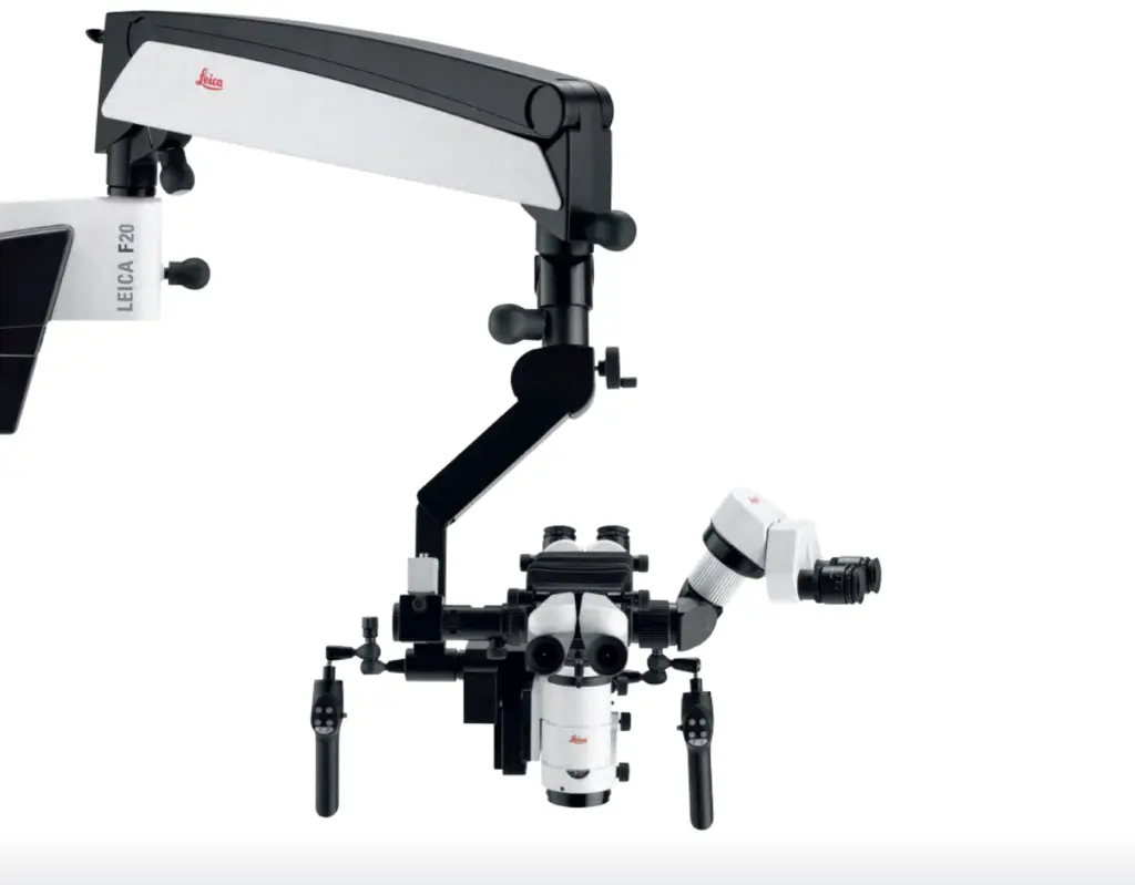
The diagnostic-operative sequence described in the literature is orderly and rational: high resolution digital radiography, operating microscope for the evaluation of the access cavity and internal details, ultrasonic tips for the targeted removal of calcified dentin under visual control, limited field CBCT when the canal is not identified. In the Buniato Studio the Leica M525 operating microscope works at magnifications up to 25x with coaxial illumination, while the Planmeca VISO G3 CBCT is used with targeted volumetry on the element of interest to obtain the position, direction and degree of obstruction of the canal. A series of cases published on Iranian Endodontic Journal formalizes this sequence and describes its usefulness for reducing the risk of iatrogenic injuries such as perforations or steps. The microscope alone, in some scenarios, is not enough: the combination with three-dimensional imaging is the step that, in the literature, has allowed previously failed treatments to be completed. The page dedicated to microscopic endodontics describes the operational flow in detail.
The role of bioceramic cements in three-dimensional closure
When the canal is made patent, the problem remains of closing it in a stable manner along its entire extension, which is often narrow and irregular. Bioceramic cements based on calcium silicates (in the literature BioRoot RCS, EndoSequence BC Sealer and evidence-based equivalents) have modified this step: they have good biocompatibility, wet the dentinal walls well and maintain volume over time, reducing the risk of voids and apical reinfection. A case report published on Dentistry Journal in 2024 describes a symptomatic calcified canal treated after a first unsuccessful attempt with the microscope alone: the integration of CBCT, optical impression, endodontic guidance and obturation single-cone with sealer bioceramic led to resolution of symptoms and radiographic success at 3 and 24 month checks. It is an example of the logic of our path, also described on the page advanced technology and state of the art: not a single solution tool, but a combination of tools that are coherent with each other.
What to expect: times, checks, maintenance
The management of a calcified canal generally requires more operating time than ordinary endodontics. There can be one or two sessions, depending on the anatomical complexity and the presence or absence of an active periapical lesion. At the end of the treatment we plan clinical and radiographic checks at a short distance and then over time: follow-ups of up to six years with clinical and radiographic resolution after integrated treatments are described in the literature. The natural tooth, once correctly treated and restored in a coronal stable manner, remains the reference standard: it avoids a prosthetic or implant process, preserves proprioception and biological architecture, and keeps future rehabilitation of the mouth predictable. Ordinary maintenance, with professional hygiene and periodic visits, is the factor that protects the result. The second opinion, in this perimeter, is not a challenge to the colleague who proposed the extraction: it is an instrumental verification before an irreversible decision.
Frequently asked questions
My canal can't be seen on x-ray: does this mean it can no longer be treated?
No. An obstruction visible on a two-dimensional radiograph does not imply, in many cases, the real absence of clinically reachable pulp tissue. The decisive evaluation is made under an operating microscope and, when the canal remains unidentifiable, by integrating a limited-field CBCT on the element. Only then can it be established whether the conservative process is truly concluded.
Does a second endodontic opinion mean rejecting what my dentist told me?
No. The second opinion is an instrumental act: it serves to verify whether imaging and direct vision change the decision-making framework, then returning a structured judgment to the patient. When the prognosis remains unfavorable we confirm the initial indication; when it is favorable we propose conservative treatment and coordinate the process with the treating colleague.
When is extraction really the right choice?
When three-dimensional imaging or clinical examination shows a documented vertical root fracture, advanced external root resorption or severe periodontal compromise of the tooth. In these cases no instrumentation significantly changes the prognosis and insisting exposes one to clinical and biological costs without an expected benefit.
For a personalized evaluation of your case, Dr. Buniato is available for one first specialist visit with complete diagnostic analysis.
Sources
- Giri K, Banga K, Arora S, Elmsmari F, Pawar AM. Management of calcified canals during root canal treatment. A systematic review of case reports. PeerJ. 2025. doi:10.7717/peerj.19900. PubMed.
- Soares de Toubes KM, Drummond de Oliveira PA, Machado SN, Pelosi V, Nunes E, Silveira FF. Clinical Approach to Pulp Canal Obliteration: A Case Series. Iran Endod J. 2017;12(4). doi:10.22037/iej.v12i4.18006. PubMed.
- Fornara R, Pisano M, Salvati G, Malvicini G, Iandolo A, Gaeta C. Management of Calcified Canals with a New Type of Endodontic Static Guide: A Case Report. Dent J (Basel). 2024;12(6):166. doi:10.3390/dj12060166. PubMed.