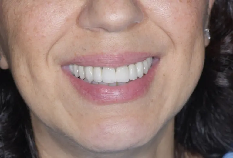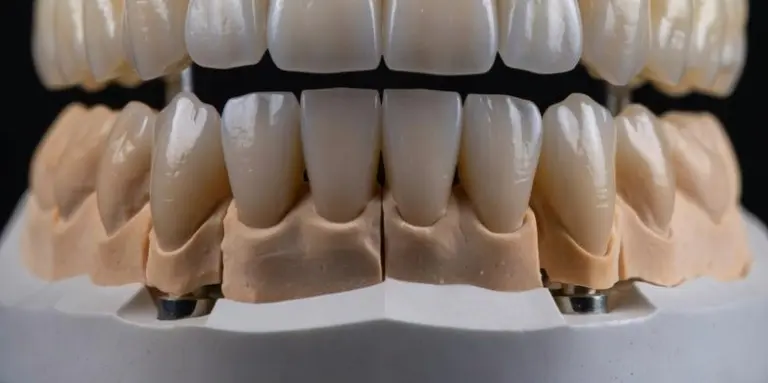
In summary. On anterior teeth, the ceramic veneer is indicated when the tooth is vital and structurally intact: the preparation generally removes 0.3 to 0.5 millimeters of enamel on the vestibular surface, a fraction of what a complete crown requires. The crown remains the correct choice when the tooth is devitalized and fragile, or when a large pre-existing reconstruction has already replaced a large part of the natural substance. The decisive criterion is not aesthetic but structural: how much healthy tooth substance remains, and how much should be sacrificed.
How much tooth substance is preserved with a veneer compared to a crown?
The difference was quantified in the laboratory. A study published in Journal of Prosthetic Dentistry measured by weight the coronal substance removed by the different preparation designs on the anterior teeth: veneer preparations remove approximately 3% to 30% of the dental crown, while full crown preparations remove 63% to 72%. In the same analysis, preparation for a metal-ceramic crown required 4.3 times more tissue removal than a veneer limited to the buccal surface.
In clinical terms, the review of the literature by Peumans and collaborators indicates for the veneer a vestibular reduction ranging from approximately 0.3 millimeters in the cervical third to approximately 0.5 millimeters in the body of the tooth, with the declared objective of remaining within the enamel. This detail is not secondary: the adhesion of the ceramic to the etched enamel is the biomechanical foundation of the veneer, and every millimeter of preserved enamel is an adhesive surface that works for the stability of the restoration. The crown, on the contrary, replaces the entire coronal surface and entrusts its retention to the geometry of the prepared abutment and the cement.
When is the veneer sufficient?
The veneer is indicated when the problem concerns the visible surface of a structurally healthy tooth. There are four typical cases: stable discolorations that do not respond to whitening, corrections of the shape or proportion of the tooth, closure of diastemas (the spaces between the front teeth), slight wear of the incisal edge or small fractures limited to the enamel.
The starting conditions matter as much as the indication. Sufficient residual enamel is needed for adhesion, a vital tooth or one that is otherwise intact in its internal structure, and a carefully evaluated occlusion. Not all cases fall within this perimeter: uncompensated bruxism, for example, documentedly increases the risk of ceramic fracture — in the study by Beier and collaborators, parafunction was associated with a 7.7 times greater risk of failure. In the presence of grinding, the veneer is not excluded a priori, but requires night protection and a more in-depth preliminary occlusal evaluation; in some paintings it is correct to orient yourself elsewhere.
When is the crown needed?
The crown is indicated when the tooth has lost, due to clinical history, the structure that would make a veneer reliable. The most frequent case is the devitalized and fragile anterior tooth: in the long-term study by Beier and collaborators, non-vital teeth restored with veneers showed a significantly higher risk of failure than vital teeth. An endodontically treated tooth, often already reconstructed and darker, benefits in many cases from the circumferential protection that only the crown offers.
The second scenario is the large pre-existing reconstruction: when extensive composite fillings have already replaced a significant part of the natural crown, the residual enamel may not be enough for predictable adhesion, and the veneer would end up sticking more to the restorative material than to the tooth. In these cases the crown is not a more invasive choice: it is the choice proportionate to a loss of substance that has already occurred. The principle remains the same in both directions — the restoration adapts to what remains of the tooth, not the other way around.
Adhesion or cementation: what really changes?
Veneer and crown do not attach to the tooth in the same way, and the difference explains much of the respective indications. The veneer has no mechanical retention of its own: it is a thin ceramic shell that becomes a single body with the tooth through adhesion - etching of the ceramic and enamel, silane, resin cement. Fradeani and collaborators, documenting a survival of 94.4% at 12 years on 182 veneers, explicitly indicate the correctness of the adhesive technique as a condition for that result. It is a protocol sensitive to details: field isolation, time management, control of each interface.
The crown follows a different logic: retention arises first of all from the geometry of the prepared stump, and cementation - adhesive or traditional depending on the material - completes it. This makes it less dependent on the presence of enamel, and is the reason why it remains reliable precisely in cases where the enamel is no longer there.
How long do ceramic veneers last? Long-term data
The longevity of veneers is among the best documented in restorative dentistry. The retrospective study by Beier and collaborators (International Journal of Prosthodontics, 2012), on 318 veneers followed up to 20 years, reports an estimated survival of 94.4% at 5 years, 93.5% at 10 years and 82.93% at 20 years; the main cause of failure was ceramic fracture. Fradeani and collaborators (2005) confirm the picture on 182 veneers in two private practices: survival of 94.4% at 12 years.
These numbers must be read for what they are: results obtained in defined conditions — correct selection of the case, conservative preparation in the enamel, rigorous adhesive technique, periodic checks. They are not a promise that can be transferred to any clinical situation; they are proof that, when the conditions are right, the veneer is a long-term restoration and not a temporary cosmetic solution.
What role does the microscope have in minimally invasive preparation?
Working in tenths of a millimeter requires seeing them. A preparation of 0.3-0.5 millimeters leaves very narrow margins of error: a few tenths more and you go beyond the enamel, exposing the dentin, with direct consequences on the quality of the adhesion. In the Buniato Studio the preparation of the veneers takes place underneath Leica M525 operating microscope, with enlargements that allow you to control the depth of reduction, the continuity of the margins and respect for the residual enamel in each phase.
The flow continues with theoptical impression via intraoral scanner 3Shape TRIOS 6, which detects the preparation margins without traditional impression materials, and ends in the dental laboratory, where the feldspar ceramic is layered by hand, layer upon layer, to reproduce the translucency of natural enamel. Instrumental precision and craftsmanship are not alternatives: the former defines the boundaries within which the latter can express itself.
Veneer or crown: synthetic comparison
| Criterion | Ceramic veneer | Complete crown |
|---|---|---|
| Tooth substance removed | Approximately 3-30% of the dental crown; vestibular reduction 0.3-0.5 mm, ideally in the enamel | Approximately 63-72% of the dental crown; reduction on all surfaces |
| Starting tooth | Vital, structurally intact, sufficient enamel | Fragile devitalized, or already extensively reconstructed |
| Typical indications | Discolorations, shape corrections, diastemas, light wear | Extended loss of substance, circumferential protection |
| Fixing | Enamel-ceramic adhesion with resin cement | Cementation on abutment with geometric retention |
| Published survival data | 93.5% at 10 years; 94.4% at 12 years; 82.93% at 20 years | High and documented; it depends on the material and clinical case |
| Main risk factors | Uncompensated bruxism, adhesion to dentin, non-vital tooth | Preparation on vital tooth: greater biological sacrifice |
Frequently asked questions
Does the veneer "ruin" the tooth?
The preparation is irreversible but conservative: it generally removes 0.3-0.5 millimeters of enamel on the vestibular surface alone, compared to the 63-72% of coronal substance required for a complete crown. The underlying tooth remains vital and structurally intact.
Can a root canal tooth receive a veneer?
In selected cases yes, but the literature documents a significantly greater risk of failure for non-vital teeth. For a fragile or already heavily reconstructed devitalized tooth, the crown is usually the most predictable choice.
Do the veneers come off easily?
No, when the adhesion occurs on enamel with a rigorous protocol: long-term studies report survivals of over 93% at 10-12 years, and the main cause of failure is not detachment but fracture of the ceramic.
I grind my teeth: can I get veneers?
Bruxism documentedly increases the risk of fracture (up to 7.7 times). It does not always exclude the veneer, but requires a preliminary occlusal evaluation and dedicated night protection; in some paintings another path is more correct.
How much enamel is needed for the veneer to be reliable?
There is no single threshold valid for each tooth: it matters how much adhesive enamel surface remains after preparation. For this reason, the evaluation is done on a case-by-case basis, with direct clinical examination and analysis of the restorative history of the tooth.
Is the preparation painful?
No: the session takes place under local anesthesia and the pain is controlled. Since these are superficial preparations, often confined to the enamel, the subsequent course is generally free of relevant symptoms.
For an evaluation of your specific case, the first specialist visit includes the complete diagnostic analysis.
Sources
- Beier US, Kapferer I, Burtscher D, Dumfahrt H. Clinical performance of porcelain laminate veneers for up to 20 years. Int J Prosthodont. 2012;25(1):79-85. PubMed.
- Fradeani M, Redemagni M, Corrado M. Porcelain laminate veneers: 6- to 12-year clinical evaluation — a retrospective study. Int J Periodontics Restorative Dent. 2005;25(1):9-17. PubMed.
- Edelhoff D, Sorensen JA. Tooth structure removal associated with various preparation designs for anterior teeth. J Prosthet Dent. 2002;87(5):503-509. doi:10.1067/mpr.2002.124094. PubMed.
- Peumans M, Van Meerbeek B, Lambrechts P, Vanherle G. Porcelain veneers: a review of the literature. J Dent. 2000;28(3):163-177. doi:10.1016/S0300-5712(99)00066-4. PubMed.