Bruxism is the repetitive activity of the masticatory muscles that leads to grinding or clenching of the teeth, and manifests itself in two distinct forms: during sleep (sleep bruxism) and when awake (waking bruxism). Those affected often do not notice it: it is the marks on the teeth and muscles that reveal it, and it is the dentist who intercepts them before the patient is aware of them. Recognizing it in time allows you to protect natural teeth and restorations from excessive load, not to "cure" a habit which, in many cases, has no definitive cure.
What is bruxism and how widespread is it?
Bruxism is a masticatory muscular activity characterized by clenching or grinding of the teeth. The 2018 international consensus does not define a disease in otherwise healthy individuals, but a behavior which, depending on the context, can represent a risk factor (for example dental wear or complications on restorations) or, in some cases, a protective factor. Distinguishing the form of sleep from that of wakefulness is important, because they have different mechanisms and management.
Prevalence data vary greatly based on the detection method. A 2019 umbrella review estimates a prevalence of waking bruxism in adults at around 22-30% and sleep bruxism at between 1% and 15%. When the diagnosis is based on polysomnography, considered the instrumental reference for sleep disorders, a population study found confirmed sleep bruxism in 5.5% of subjects. The wide range depends on the fact that questionnaires, clinical examination and instrumental recordings measure different things.
What are the signs that the dentist detects?
The first sign is wear: flat, shiny wear faces on the occlusal surfaces and incisal edges, with flattening enamel and, in advanced cases, exposed dentin. Added to this are cracks in the enamel, microfractures, chipping of the margins, increased sensitivity to cold and chewing, mobility of individual elements and, on restorations, detachments or recurrent fractures. They are signs that the patient rarely connects with each other, but which in the clinical examination make up a coherent picture.
Alongside the dental signs there are muscular and joint ones. Pain or fatigue in the masticatory muscles, particularly upon awakening, morning tension-type headache, noises or pain in the temporomandibular joint, hypertrophy of the masseter muscles and dental impressions on the mucosa of the cheeks are elements that lead to suspicion. The literature describes a plausible association between bruxism and signs and symptoms of temporomandibular disorders, which is why the examination is not limited to the teeth but includes muscles and joints.
What consequences does it have on natural teeth and restorations?
On natural teeth, the repeated load of bruxism contributes to the wear of surfaces, the appearance of cracks and, over time, to the reduction of the height of the clinical crowns with possible involvement of sensitivity and aesthetics. It is important to point out that the role of bruxism in dental wear is considered one factor among others, not necessarily the main cause: acidic diet, habits and other elements contribute to the picture.
On restorations the implications are more defined. A critical review dedicated to bruxism and prosthetic rehabilitation concludes that bruxism is associated with an increase in mechanical and technical complications of prosthetic products - for example ceramic fractures or chipping of veneers and crowns - although it was not, in that analysis, associated with a reduction in implant survival. The 2019 umbrella review also reports possible biomechanical complications affecting the implants, maintaining caution on the conclusions. The practical consequence is that, in a person with bruxism, the design of veneers, crowns and rehabilitations takes into account the expected occlusal load, on the materials and on the contact pattern, to reduce the risk of early complications.
How is bruxism diagnosed?
The diagnosis starts from the clinical examination: evaluation of the wear faces, existing restorations, the masticatory muscles and the temporomandibular joint, together with the medical history (headache upon awakening, reported grinding by those sharing the sleep, periods of stress). This allows us to distinguish active wear from previous and stabilized wear, a fact that changes subsequent decisions.
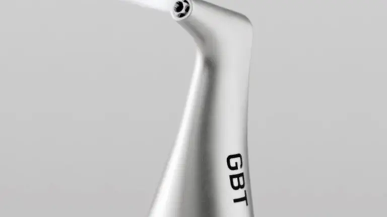
When you need to objectively document occlusion and dental contacts, digital analysis is of support. In the Studio Buniato the 3Shape TRIOS 6 intraoral scanner acquires aoptical impression of the arches and occlusal relationships, useful for mapping wear surfaces, recording a reference over time and precisely designing a possible device. The diagnosis remains clinical: the instrumental recording of sleep bruxism (polysomnography, electromyography) belongs to specialized sleep contexts and is reserved for cases in which the situation requires it.
How is it managed? The custom-made bite and other sizes
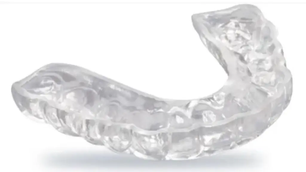
The most consolidated measure is the custom-made occlusal bite, a rigid device that is placed between the arches. Its documented role is to protect teeth and restorations from the load, distributing forces and absorbing friction which otherwise acts directly on the enamel and ceramic. It is useful to be precise about what the bite does and what it does not do: a Cochrane review on occlusal splints in sleep bruxism found insufficient evidence to demonstrate that the device reduces bruxism activity in itself compared to other interventions, even without adverse effects. The bite, therefore, does not "turn off" bruxism: it protects the structures while the habit persists.
The creation of the bite can follow a digital flow: from the optical impression with scanner the device is designed on the computer and it is produced with 3D printing (in the study the SprintRay MIDAS printer), with adaptation and control of contacts in the mouth. This is accompanied by behavioral and sleep hygiene measures - regularity of hours, reduction of caffeine, alcohol and tobacco in the evening hours, stress management - consistent with the factors that the literature most consistently associates with bruxism. None of these measures are promises of resolution: they are risk control tools.
When is a gnathological approach needed?
A structured gnathological approach is indicated when bruxism is associated with persistent muscle or joint pain, limitation of mandibular movements, symptomatic joint noises or wear that has already altered the occlusal relationships and requires rehabilitation. In these cases the evaluation integrates teeth, muscles and temporomandibular joint, and the therapeutic sequence is defined before any reconstructive intervention, so that any veneers or crowns are designed on a stable occlusal scheme.
Not all cases require this level. Many situations can be managed with night protection and wear monitoring over time. The guiding criterion is the presence of symptoms and documented progressive damage, not the simple finding of grinding: interpreting this distinction correctly avoids both undertreatment and disproportionate interventions.
Frequently asked questions
How do I know if I grind my teeth in my sleep?
Often the first clue comes from the dentist, who recognizes signs of wear, cracks or sensitivity during the clinical examination. Other signs include tension-type headache upon waking, fatigue in the chewing muscles in the morning and reports of grinding noises from those sharing the sleep.
Is the bite really necessary or is it just a temporary remedy?
The bite has a documented role in protecting teeth and restorations from occlusal loading. However, the available evidence does not demonstrate that it reduces bruxism activity itself: it does not eliminate the habit, but limits the damage it produces on the structures. For this reason it should be understood as a protective measure to be maintained over time, not as a definitive cure.
Does bruxism ruin veneers and crowns?
Bruxism is associated with an increase in mechanical complications of restorations, such as fractures or chipping of the ceramic. It does not mean that rehabilitation is inadvisable, but that the design takes into account the expected load, in the choice of materials and in the contact pattern, and that night protection is often part of the plan.
Is stress the cause of bruxism?
The origin of bruxism is multifactorial. Among the factors most consistently associated, the literature indicates the use of alcohol, caffeine and tobacco and some drugs; stress is among the elements considered, but it is not a single cause. Acting on these factors is a control measure, not a guarantee of resolution.
Do I need to do a sleep test for diagnosis?
In most cases, no. The diagnosis is clinical and is based on the examination of teeth, muscles and joints, integrated when useful by digital analysis of the occlusion. Instrumental sleep recording is reserved for specific situations, in dedicated specialist contexts.
Sources
- Lobbezoo F, Ahlberg J, Raphael KG, et al. International consensus on the assessment of bruxism: Report of a work in progress. J Oral Rehabil. 2018;45(11):837-844. doi:10.1111/joor.12663. PubMed.
- Melo G, Duarte J, Pauletto P, et al. Bruxism: An umbrella review of systematic reviews. J Oral Rehabil. 2019;46(7):666-690. doi:10.1111/joor.12801. PubMed.
- Manfredini D, Winocur E, Guarda-Nardini L, Paesani D, Lobbezoo F. Epidemiology of bruxism in adults: a systematic review of the literature. J Orofac Pain. 2013;27(2):99-110. doi:10.11607/jop.921. PubMed.
- Maluly M, Andersen ML, Dal-Fabbro C, et al. Polysomnographic study of the prevalence of sleep bruxism in a population sample. J Dent Res. 2013;92(7 Suppl):97S-103S. doi:10.1177/0022034513484328. PubMed.
- Johansson A, Omar R, Carlsson GE. Bruxism and prosthetic treatment: a critical review. J Prosthodont Res. 2011;55(3):127-136. doi:10.1016/j.jpor.2011.02.004. PubMed.
- Macedo CR, Silva AB, Machado MA, Saconato H, Prado GF. Occlusal splints for treating sleep bruxism (tooth grinding). Cochrane Database Syst Rev. 2007;(4):CD005514. doi:10.1002/14651858.CD005514.pub2. PubMed.
For an evaluation of your specific case, the first specialist visit includes the complete diagnostic analysis.