The CBCT before removing a wisdom tooth is not a routine examination: it is needed when the root of the tooth approaches the nerve of the jaw or the maxillary sinuses. Without these signals, a well-read overview remains sufficient to plan the extraction.
For whom CBCT on wisdom teeth may be useful
Three-dimensional diagnosis with CBCT is indicated for adult patients with impacted wisdom teeth (third molars) or in disodontiasis, especially lower ones, when the root may be in close relationship with the nerve of the mandible, called the inferior alveolar canal. These are generally patients who have already performed an overview and in which the two-dimensional image leaves a precise doubt: where exactly does the nerve pass and how close is it to the root of the tooth to be removed?
The examination is also particularly useful when the lower wisdom tooth shows signs of resorption of the adjacent second molar, distal bone loss, or when the planning includes the option of coronectomy, i.e. the removal of only the crown with preservation of the roots to protect the nerve. In these cases an evaluation with advanced technology is an integral part of the route.
When the overview is already enough
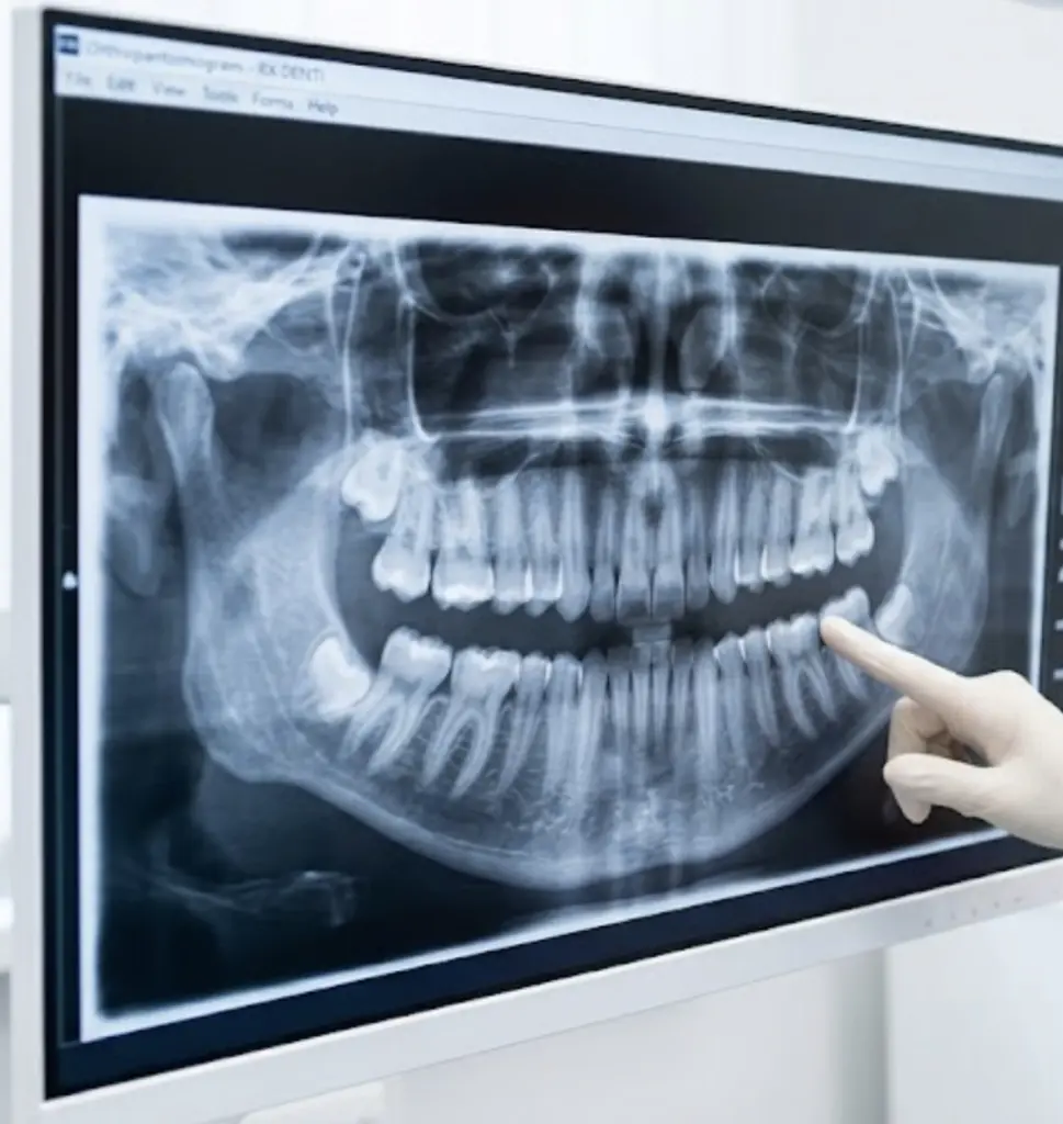
Not all wisdom teeth require a three-dimensional examination. On the panoramic view, if the roots appear clearly separated from the mandibular canal and no critical overlaps are observed, the basic examination is already adequate to set up the extraction. The same applies to upper wisdom teeth without complex relationships with the maxillary sinus visible in two dimensions, and to cases of partial eruption without deep bone inclusion.
The operating principle is clear: CBCT is justified when it can change the clinical decision. If the overview already provides useful information, adding a three-dimensional examination is not beneficial and only increases the radiation dose. This is why in the practice we prescribe CBCT only after reading the signals on the overview and relating them to the overall clinical picture.
Radiographic signals that raise the priority of CBCT
The literature has identified some signals on the overview that are significantly associated with the absence of corticalization between the root of the wisdom tooth and the inferior alveolar canal on CBCT. These signs, described by Rood and Shehab, are six:
- interruption of the cortical white line of the canal;
- darkening of the root at the canal;
- darkening of the channel itself;
- root deviation;
- channel narrowing;
- channel deviation.
The presence of one or more of these signs makes the investigation with CBCT clinically justified. Their absence suggests a separate report and suggests proceeding with the overview only. It is the filter that avoids the overprescription of three-dimensional exams and recognizes the cases in which an in-depth view really changes the surgical plan.
As we decide in the Buniato Study
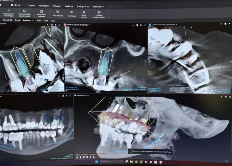
The sequence in the study is always the same. First there first specialist visit with anamnesis and overview analysis already available or performed from scratch. Then the evaluation of the radiographic signals described above. Only in case of actual indication, the CBCT acquisition with our Planmeca equipment, on a volume targeted to the area of interest to contain the radiation dose.
The report is read together with the patient, showing where the jaw nerve runs and how it relates to the roots of the tooth to be removed. When proximity risk is high, we openly discuss surgical alternatives, including coronectomy, and postoperative management scenarios. The decision takes into account the clinical risk, age and symptoms: CBCT does not automate the choice, it makes it more transparent and shared.
What changes in the surgical plan after the 3D exam
CBCT does not amplify risk: it stratifies it. A 2025 study of 115 lower wisdom teeth observed that, in the presence of a cortical defect of the lower alveolar canal visible on CBCT, intraoperative exposure of the nerve occurred in 73.9% of cases. The data does not mean that the injury is certain: it means that CBCT precisely identifies high-risk situations and allows you to prepare technically in a different way. The length of the cortical defect visible in 3D was correlated with nerve exposure, with a odds ratio of 1.38 for every additional 0.1 mm.
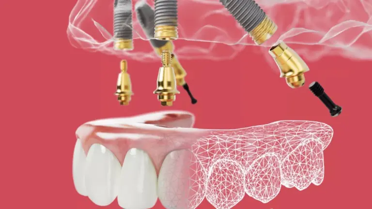
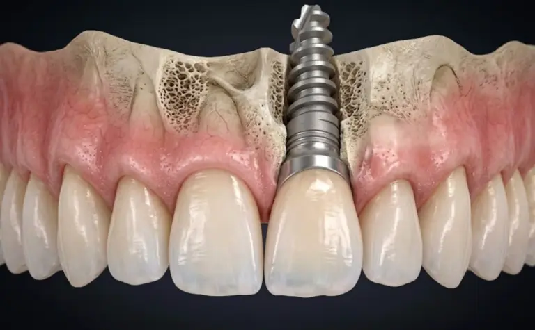
In practice, after an informative CBCT the plan can change in three directions: standard extraction with specific surgical precautions, targeted fragmentation of the root during surgery, or coronectomy with preservation of the roots when complete extraction would expose the nerve. The same logic applies when the 3D data is used to set a implant rehabilitation in the same region.
What risks does three-dimensional planning reduce
The main risks of lower wisdom tooth surgery are injury to the lower alveolar nerve, with alteration of sensitivity of the lip and chin, oro-antral communication for the upper wisdom teeth in close relationship with the maxillary sinuses, and post-extraction complications resulting from approximate planning.
A planning that integrates well-interpreted panoramic and targeted CBCT, when indicated, concretely reduces these risks: the surgeon knows in advance the precise point of proximity between the root and the nerve, chooses the least invasive technique possible and prepares instruments and operating times in a coherent manner. When the relationship is critical, he may propose a coronectomy instead of complete extraction. The approach does not guarantee zero risk, but takes the decision out of improvisation.
What to expect during the exam and afterwards
CBCT acquisition with Planmeca equipment requires a few seconds of scanning, standing, without contrast medium and with a field of view limited to the affected region, to keep the radiation dose proportionate to the clinical question. The exam is outpatient and immediately reportable: the patient does not add days of waiting to the procedure and can review the 3D image together with the clinician in the same session. The report enters directly into the surgical plan discussed in the office, with the same logic of precision applied in the treatments carried out at operating microscope.
In the course of time, a well-established three-dimensional planning reduces intraoperative variability, limits surgical times and makes post-extraction management more predictable. An extraction decided today on the basis of solid data limits neurological sequelae and surgical revisions, and protects the health of the adjacent teeth, in particular the second molar.
Frequently asked questions
I've already done the overview, why add the CBCT?
The overview remains the starting point. CBCT is added only when the overview shows precise signals of proximity to the nerve or when other clinical elements require it. If there is no indication, it is not performed: the objective is to prescribe the test when it can change the surgical plan, not as an automatic step.
Does CBCT expose you to more radiation than panoramic?
CBCT involves a higher dose than panoramic, but is performed with a field of view limited to the area to be investigated. For this reason it is prescribed only when the diagnostic benefit is clear and proportionate. The selection of volume and acquisition parameters is an integral part of the clinical decision.
What is a coronectomy and when is it offered?
Coronectomy consists in removing only the crown of the wisdom tooth, leaving the roots in place, when these are too close to the nerve of the jaw. It is offered in selected cases, after a CBCT that documents the risk and after explicit informed consent that clarifies limits and follow-up.
Are the exam and the extraction done on the same day?
The CBCT is a short, outpatient examination, which can also be performed before the operating session. The extraction is scheduled in a dedicated session. When the risk is high it is important not to compress the two phases into a single step and to provide a moment for shared reading of the report.
Can the risk to the jaw nerve be completely avoided?
No technique eliminates risk absolutely. However, a planning that integrates overview, clinical evaluation and targeted CBCT allows you to manage the risk in an informed way and to choose the most conservative surgical strategy for the individual case, reducing the probability of permanent sequelae.
For a personalized assessment of your case, Dr. Buniato is available for an initial specialist visit with complete diagnostic analysis.
Sources
- Patel PS, Shah JS, Dudhia BB, Butala PB, Jani YV, Macwan RS. Comparison of panoramic radiograph and cone beam computed tomography findings for impacted mandibular third molar root and inferior alveolar nerve canal relation. Indian J Dent Res. 2020. DOI: 10.4103/ijdr.IJDR_540_18. PMID: 32246689.
- Barraclough J, Power A, Pattni A. Treatment Planning for Mandibular Third Molars. Dent Update. 2017;44(3):221. DOI: 10.12968/denu.2017.44.3.221. PMID: 29172329.
- Chen K, Chen Y, Chen P, Jiang J, Wang E, Guo C. Is cone beam computed tomography accurate in predicting inferior alveolar nerve exposure during mandibular third molar extraction? BMC Oral Health. 2025. DOI: 10.1186/s12903-025-05716-w. PMID: 40055692.
- Leung YY, Hung KF, Li DTS, Yeung AWK. Application of Cone Beam Computed Tomography in Risk Assessment of Lower Third Molar Surgery. Diagnostics (Basel). 2023;13(5):919. DOI: 10.3390/diagnostics13050919. PMID: 36900063.
- Weckx A, Agbaje JO, Sun Y, Jacobs R, Politis C. Visualization techniques of the inferior alveolar nerve (IAN): a narrative review. Surg Radiol Anat. 2016;38(1). DOI: 10.1007/s00276-015-1538-0. PMID: 26163825.